
Resources
 Part of the Oxford Instruments Group
Expand
Collapse
 Part of the Oxford Instruments Group
The Dragonfly provides high-quality confocal images across scales, from the subcellular level to entire organisms, and significantly increases productivity. It combines speed and sensitivity, enabling researchers to observe dynamic events and live organisms for extended periods. The B-TIRF and super-resolution modules reveal finer details, such as viral infection dynamics and chromatin or organelle ultrastructure.
Each Dragonfly high-speed confocal includes Fusion software for effortless image acquisition and Imaris, the market leader in image analysis software.The Dragonfly 600 is the flagship model. It delivers a full-featured system and allows for effortless cross-scale imaging.
Discover our NEW Dragonfly 400 System
Deeper Imaging. Deeper Insights. Deeper Understanding.
Dragonfly spinning disk confocal delivers leading performance for industrial and academic users. Its modular design adds functionality and capability at each level.
Use the hotspots below to explore the key features and benefits of Dragonfly.
Whether imaging glial cells, axonal transport, large tissue sections, expanded samples (Expansion Microscopy), or super-resolved dendritic spines (SMLM), Dragonfly is the instrument of choice.

The high sensitivity, low phototoxicity/ photobleaching and speed are ideal features for live cell imaging applications such as microtubule dynamics or intracellular organelle trafficking
With B-TIRF researchers can address live events at the edge of the coverslip, such as vesicle fusion or membrane receptor trafficking. B-TIRF delivers increased signal to noise in SMLM applications
The HLE can be used in combination with any imaging modality (confocal, widefield and B-TIRF) for SMLM applications, such as ultrastructural organisation of organelles, chromatin and epigenetic organisation, mitochondrial membrane structure.

Dragonfly allows researchers to study cancer cell behaviour interaction with the environment and spatial distribution of the tumor both in fixed and live cells.

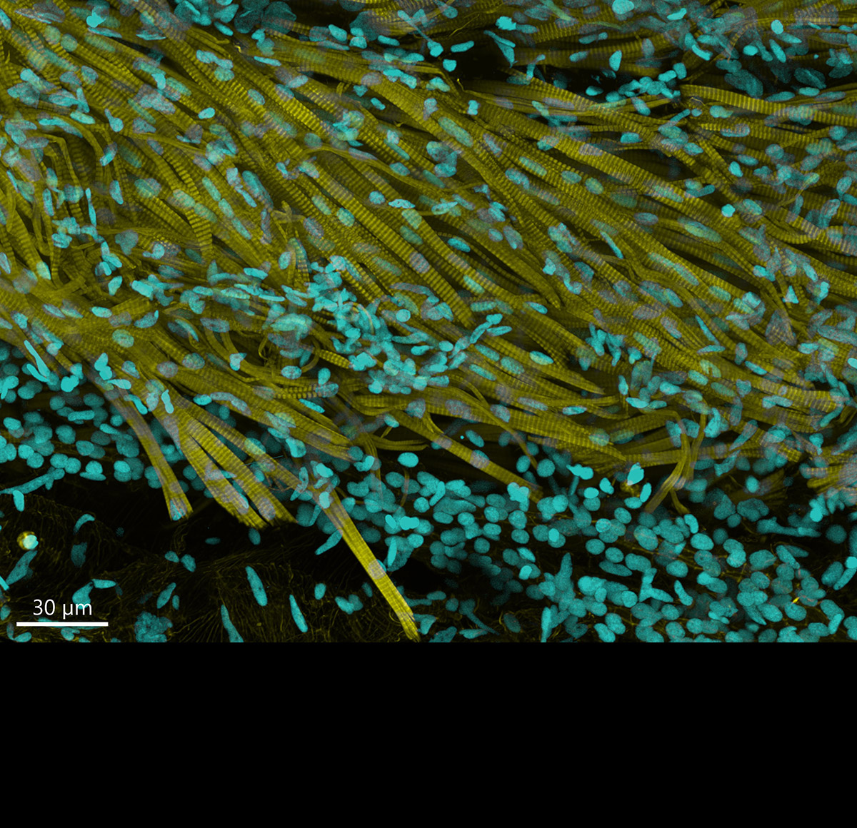
Image credit: Julien Rességuier at NorMIC (University of Oslo)
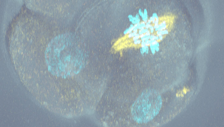
Dragonfly's optimized pinhole size allows imaging very deep into thick organisms - up to the millimetre range.

Omics research is used for multiple applications, such as predicting disease progression based on gene expression mappings, assessing tumor microenvironments and cancer severity, etc.
Omics-related sciences gather information about Xn biological molecules to characterize and quantify entire pools of molecules.
Dragonfly's highly sensitive detectors, exceptionally high background rejection, acquisition speed and Uniform illumination make it the perfect system for Omics applications

The resolution and sensitivity delivered by Dragonfly 600 makes it a unique system for studying micro-organisms:


Dragonfly performs at a very high level and its ease of use allows researchers to acquire valuable data at speed. The system really excels for applications that are more demanding, such as high resolution live cell imaging.
Derek Toomre, PhD - Professor of Cell Biology at Yale School of Medicine.
At its core, Dragonfly, is a multi-point confocal for high-speed and high-sensitivity imaging.
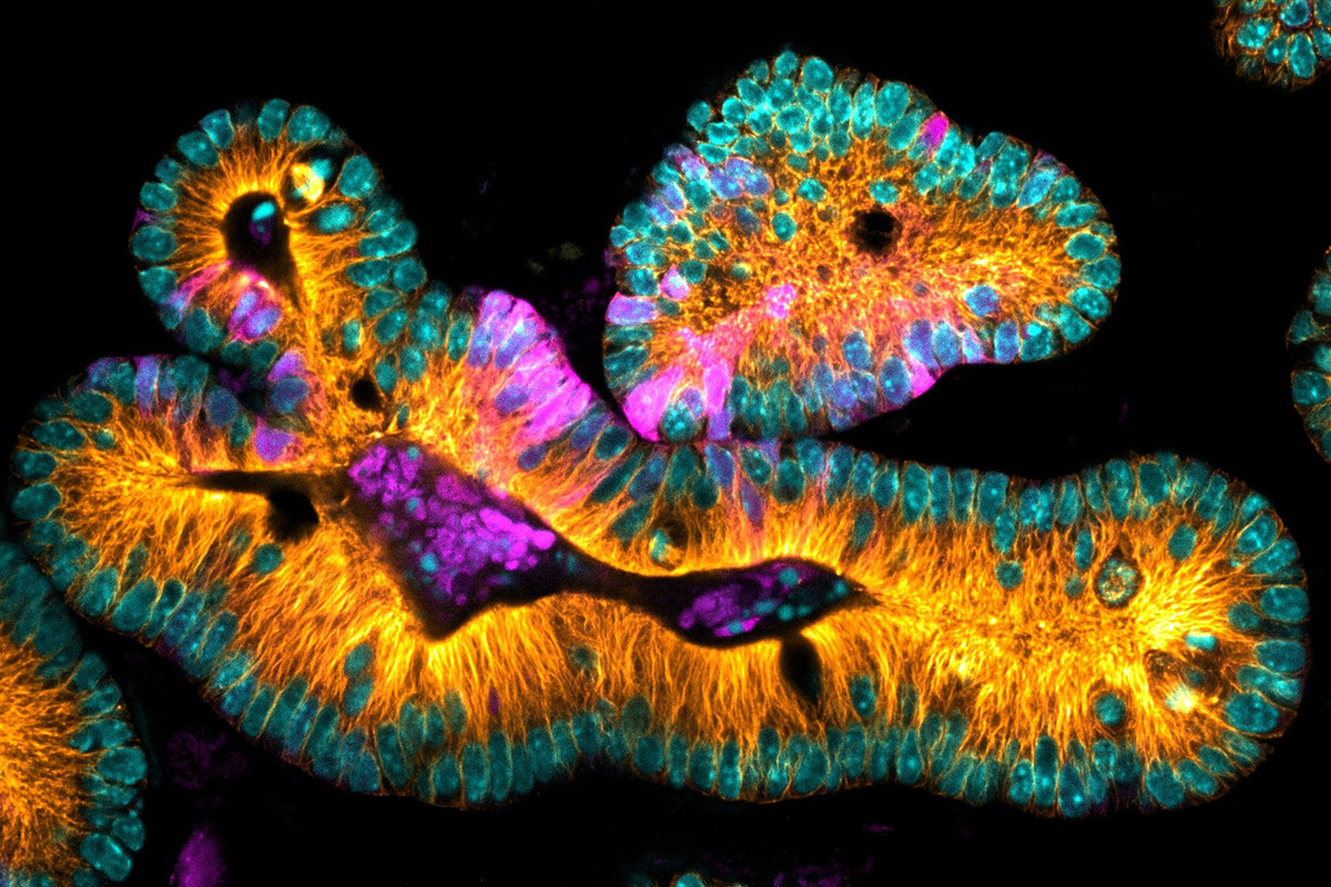
 Sample courtesy: Ching-Ho Chang, PhD; Harmit Malik Lab – Fred Hutchinson Cancer Research Center")
Borealis Total Internal Reflection Fluorescence (B-TIRF) is Andor’s patented TIRF module available on Dragonfly 600. Applications include a) Image dynamic events at the edge of the coverslip (e.g. membrane fusion events, receptor signalling, and infection dynamics) or b) Increasing the signal-to-background ratio in SMLM imaging.
With Dragonfly you can select from a number of super-resolution approaches all from the same imaging system. Resolutions down to 6 nm can be achieved.

SRRF (Super-Resolution Radial Fluctuations) offers a highly effective, one-click, software-based approach to super-resolution imaging. Our exclusive development, SRRF-Stream+, accelerates processing by delivering real-time super-resolution.


Laser-illuminated widefield fluorescence is ideal for studies that do not benefit from confocal imaging. Dragonfly users can easily change to widefield mode, with no need to switch microscope ports.


All imaging modalities can be combined with transmitted light techniques (DIC or phase contrast) to expand your imaging capabilities even further. DIC or Phase Contrast can be used in a variety of ways including:


Dragonfly is fast, sensitive, and fantastically flexible. We have 120 research groups working at our Core Facility every year, and they bring in everything you could imagine to image. Having the flexibility to switch between different modes and colors, and having a system that is very sensitive for single-molecule imaging through to spinning-disk confocal microscopy, is amazing for us.
Prof. Jeffrey Caplan - Director of Bioimaging and Associate Professor, University of Delaware.
Fusion is designed to meet the requirements of today’s researchers, providing ease of use and immediate visual feedback for data review.

We have worked with Picasso developers to allow Imaris .ims files to be read in Picasso. Users have the flexibility to go to open-source software to retrieve SMLM data and 3D SMLM localizations.
3D rendering and statistical measurements are then delivered in Imaris. Please read our article on the acquisition to analysis workflow.

Dragonfly models come with the introductory Imaris Quant package for users to:
Additional Imaris modules are also available including tools for tracking and reporting motility metrics, quantifying cells and their intracellular organelles, and tracing neurons for reporting their specific characteristics.

Use microscope selector tool below to have an overview of the best Dragonfly for your application or technique
Filter by:
A flexible, multi-modal platform that offers confocal, laser based widefield, transmitted light and SRRF-Stream+ functionality in one system.

3D SMLM widefield imaging capabilities (DNA-PAINT). Spatial transcriptomics / large model organisms high-performance capabilities.

Fully configured Dragonfly system, adding 3D SMLM in widefield, confocal, B-TIRF (DNA-PAINT, dSTORM and PALM) and live cell TIRF imaging.

All Dragonfly imaging systems are compatible with Andor Photostimulation products. Please note that we indicate the best system for your application. In some cases, a lower model could deliver acceptable results. Please see our technical article selecting your Andor Microscope for more information.


 Powered by Bioz
 Powered by Bioz